Figure 3.
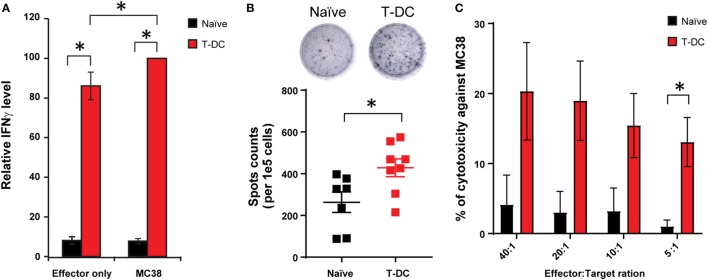
Activation of tertiary lymphoid structure (TLS)-residing lymphocytes by MC38 tumor lysate-pulsed DC (T-DC) immunization. (A) DCs were isolated from mouse bone marrow and pulsed with MC38 tumor lysate. 1e6 T-DCs were injected subcutaneously into TLS-bearing mice once a week for 3 weeks. T cells were isolated from the TLSs of mice immunized with T-DC vaccines or naïve mice, and incubated in medium alone (effector only group) or with irradiated MC38 cells (MC38 group) for 24 and 48 h. Supernatants were collected and tested for IFNγ levels using ELISA kits. IFNγ levels were normalized to the group of T-DC samples incubated with MC38 cells (n = 19–21 for naïve group, n = 12–15 for T-DC group). (B) Detection of IFNγ secretion by purified T cells from the TLSs. Representative wells were seeded with 1.25e5 cells from indicated groups. Spots were enumerated and normalized to cell number (n = 7 for naïve group, n = 8 for T-DC group). (C) Isolated TLS-residing T cells (effector cells) were incubated with labeled MC38 cells (target cells) at indicated ratio. Released chromium-51 was collected and measured after 5 h incubation (n = 3 for naïve group, n = 5 for T-DC group). Data are presented as mean ± SE. *p < 0.05.

