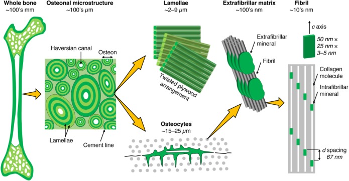
Figure 1.
Bone consists of either a porous trabecular framework or a dense cortical structure. In cortical bone (e.g., in the middiaphysis of the femur), the microstructure consists of osteons (170- to 250-µm diameter), which are the units of bone produced during remodeling. Osteons contain a central vascular canal, the Haversian canal (60- to 90-μm diameter), concentrically surrounded by lamellae having a twisted plywood arrangement, where neighboring lamellae have different fibril orientations. Osteocytes reside in lacunae interconnected through canaliculi (100- to 400-nm diameter). Lamellae are composed of collagen fibrils (80- to 100-nm diameter). Fibrils are surrounded by extrafibrillar mineral platelets. Within the fibrils, type I collagen molecules and carbonated apatite crystallites form a nanocomposite structure.

