Figure 3.
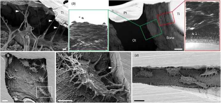
Direct attachment of osteocytes to the implant surface. (a) Osteocytes (Ot) retain connectivity to the implant surface after 4 y of clinical function through canaliculi (arrowheads). (b) Ultrastructural similarities exist between the bone-implant interface and the bone-osteocyte interface (high-angle annular dark-field scanning transmission electron microscopy and electron tomography), where both comprise highly aligned collagen fibrils forming typical rope-like bundles. Scale bar: 1 µm. (Adapted with permission from Shah et al. 2015. Copyright 2015, American Chemical Society.) (c) Interconnected osteocyte lacuno-canalicular network within 60-µm-wide features on the surface of 3D printed Ti6Al4V. (d) An osteocyte (box in c) attaches to the implant surface through numerous branching canaliculi (one of which is indicated by an arrowhead). (e) Interconnected osteocytes within a 14-µm-wide crevice on the surface of 3D printed Ti6Al4V. (Adapted with permission from Shah, Snis, et al. 2016. Copyright 2016, Elsevier.) Scale bars: 10 µm (c, e), 5 µm (d). Pt, platinum; Ti, titanium.

