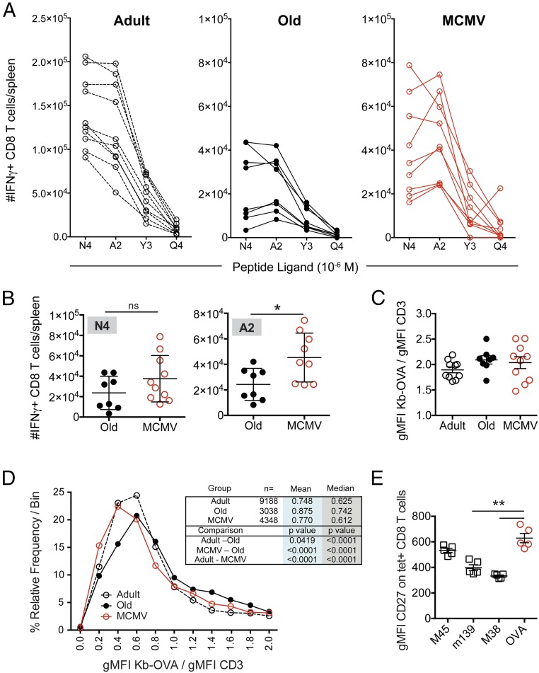
Fig. 4.
OVA-specific cells in MCMV+ mice show altered avidity for cognate antigen. (A) On day 7 after Lm-OVA infection, splenocytes were stimulated with SIINFEKL (N4) peptide or three different altered peptide ligands (A2, Y3, and Q4) at 10−6 M. The number of cells able to produce IFN-γ in response to each was assessed by FCM. Data show the individual responses for adult (open circles, n = 10) and old animals (black circles, n = 8), or MCMV+ mice (red circles, n = 9). (B) Comparison of the number of IFN-γ+ cells responding to N4 (Left) or A2 (Right) peptides in old vs. MCMV+ old mice. Significance determined by Mann–Whitney U test. (C) The gMFI of Kb-OVA257–264 tetramer binding normalized to total CD3 staining. Data depict mean ± SEM. (D) The distribution of binned Kb-OVA/CD3 gMFI of individual cells (as a measurement of TCR avidity) for adult (open, dashed line), old (black), and MCMV+ old (red) mice. (E) Splenocytes from MCMV+ old mice were stained with tetramers against three MCMV epitopes (M45, noninflating; M38 and m139, inflating), as well as the Kb-OVA257–264 tetramer. The gMFI of CD27 on each tetramer+ population was assessed. *P < 0.05, **P < 0.01. For all analyses, data were pooled from two independent experiments with n = 4–5 mice/experiment.

