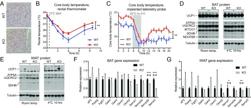
Fig. 4.
PM20D1 mice maintain a higher body temperature following cold exposure. (A) Representative BAT sections stained with H&E from WT and PM20D1-KO mice. (Magnification: 10×.) (B and C) Core body temperatures at room temperature and after transfer to 4 °C as measured by rectal thermometer (B) or telemetry probe implanted into the i.p. cavity (C) in WT and PM20D1-KO mice. Immediate transition from room temperature to 4 °C is indicated by the dashed gray line (B), and a slow transition over 3 h from room temperature to 4 °C is indicated by the gray bar (C). (D and E) Protein levels of UCP1, mitochondrial proteins, and tubulin in BAT (D) or iWAT (E) of WT and PM20D1-KO mice at room temperature or after 10 h at 4 °C. (F and G) mRNA levels of the indicated genes in WT and PM20D1-KO mice from BAT (F) or iWAT (G) at room temperature. All mice were males, 8–12 wk of age. Data are shown as means ± SEM; *P < 0.05, **P < 0.01, ***P < 0.001. For B and C, n = 6–12 per group; for F and G, n = 5–6 per group.

