Figure 2.
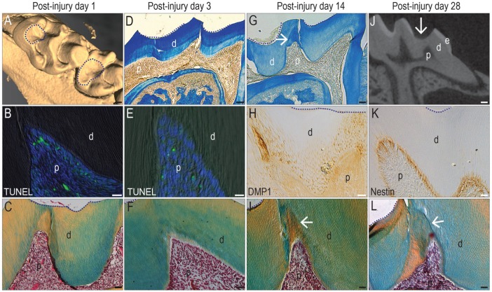
A dentin injury model elicits a reparative response from odontoblasts. In adult maxillary molars, µCT imaging identifies sites of (A) enamel and some dentin removal but no pulp exposure (dotted circles). On PID1, (B) representative tissue sections stained with TUNEL show evidence of apoptosis. (C) On an adjacent pentachrome-stained tissue section, the pulp exhibits a normal morphology (in all panels, dotted line indicates the edge of the injury). On PID3, (D) aniline blue staining indicates a dentin bridge between the injury (dotted lines) and the pulp. (E) On an adjacent tissue section, TUNEL identifies few apoptotic cells, and (F) the pulp continues to exhibit a normal morphology. By PID14, (G) aniline blue staining identifies new dentin formation (arrow) underneath the injury site (dotted lines), and (H) DMP1 immunostaining identifies odontoblasts responsible for secreting the new matrix and (arrow, I) new dentin formed. By PID28, (J) µCT sections show that a dentin bridge separates the injury site from the pulp (arrow). (K) Odontoblasts underneath the injury site are nestin+ve, and (L) the new dentin formed by the surviving odontoblasts has protected the pulp from exposure (arrow). d, dentin; e, enamel; p, pulp; PID, postinjury day; µCT, micro–computed tomography. Scale bars: 100 µm (A, D, G, J), 25 µm (B, C, E, F, H, I, K, L).

