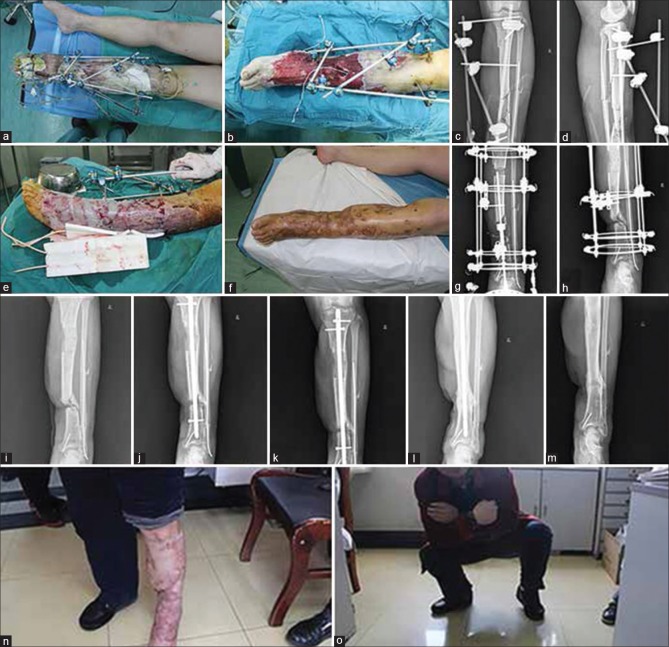
Figure 1.
(a) Clinical photograph of a patient (male, 48-year-old) who was injured in a car accident. Seven days after the emergency debridement surgery, the wound showing obvious infection (b) Clinical photograph of same patient showing that after repeated debridement, soft tissue damage and bone defect, were obvious (c and d) X-ray anteroposterior and lateral views of both bones leg showing that the external fixator was used to fix the fractures in emergency (e) Clinical photograph showing that after the wound infection was controlled, we covered the wound with a free skin graft (f) Clinical photograph showing that wound infection was in control, and the skin graft survived (g) X-ray anteroposterior view of leg bones showing that the Ilizarov circular external fixation frame was used in bone transport to treat bone defects (h) X-ray anteroposterior view of leg bones showing that the defect of the bone was about 8 cm in size, and nonunion of the bone was observed (i) The Ilizarov circular external fixation frame was removed after the bone transport was completed (j) We fixed the fracture with an intramedullary nail and the iliac bone graft was done (k) The bone callus showed obvious growth at 3 months after the internal fixation surgery (l) The fracture healed well 8 months after the internal fixation surgery (m) The internal fixation was removed 1 year after the internal fixation surgery (n) The appearance of the limb was satisfactory, and the patient resumed normal life within 14 months after the procedures (o) Clinical photograph showing that limb function was good and satisfactory

