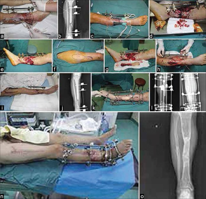
Figure 2.
(a) Clinical photograph showing that the patient (male, 26-year-old) was injured in a car accident. Seven days after the emergency debridement surgery, the wound shows obvious infection with a considerable amount of purulent secretion (b) X-ray leg bones anteroposterior view showing that the external fixator was used to fix the fractures in emergency cases, and the tibia was crushed severely. (c) Clinical photograph showing that after several rounds of debridement, wound was covered using vacuum-closed drainage. (d) After repeated debridement, we removed a lot of necrotic bone and infected soft tissue. (e) Clinical photograph showing that after repeated debridement, the infection was eliminated. (f) Clinical photograph showing that after we designed a free anterolateral thigh composite tissue flap to cover the wound. (g) Clinical photograph showing that the composite tissue flap was prepared. (h) Clinical photograph showing that after flap crossed the subcutaneous tunnel to cover the multiple irregular wounds. (i) Clinical photograph showing that the infection was controlled, and the tissue flap and skin graft survived. (j) X-ray anteroposterior view of leg bones showing that the tibial defect was about 8 cm in size. (k) Clinical photograph showing that the Ilizarov circular external fixation frame was used for bone transport to treat the bone defect. (l) X-ray anteroposterior view of leg bones showing that the Ilizarov circular external fixation frame (m) X-ray lateral view of leg bones showing the callus at 3 months after the bone transport procedure (n) Clinical photograph showing the lower limb appeared well at 1 year after the procedures. (o) X-ray of leg bones anteroposterior view at 1 year followup showing that fracture healed well

