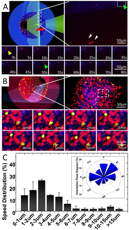
Figure 5.

Real-time imaging of monocyte perfusion, adhesion and migration inside TEBV. (A) Monocyte perfusion and adhesion in TEBV. Upper left panel, a schematic illustration of the imaging focal-plane. Upper right panel, a full view of monocyte perfusion in TEBV. Yellow arrow: a monocyte adhered to endothelium during its transmigration. Green arrow: a new monocyte attachment event. White arrow: monocyte in perfusion. Lower panel, montage of the monocyte attachment and transmigration events (Supplemental Movie 2). (B) Monocyte migration on the TEBV endothelium. Upper left panel, a schematic illustration of the imaging focal-plane. Upper-right panel, a full view of monocyte migration on TEBV endothelium. Lower panel, montage of leukocyte migration on TEBV endothelium (Supplemental Movie 3). (C) Quantification of leukocyte migration speed distribution on TEBV endothelium and their migration directionality with Rose Diagram (inserted panel). The flow direction is set as degree zero. Results were calculated from 60 leukocytes in three experiments. Data are shown as mean ± SEM.
