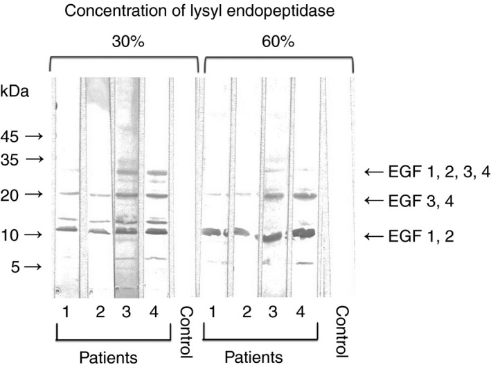
Figure 4.

Representative experiment showing patient autoantibody binding to EGF‐like domains of PS. SDS‐PAGE was performed using a polyacrylamide gradient gel (5‐20%). For the preparation of fragments of EGF‐like domains in PS, PS was incubated with high concentrations (30% or 60%) of lysyl endopeptidase (cleaves COOH‐terminal of lysines) at 37°C for 1 h in the presence of Ca2+ (2 mmol L−1). PS (3 μL of 600 μg/mL solution) was applied to each lane. Transfer to PVDF membrane was done for 20 minute at 0.1 amps. Membranes were blocked for 1.5 hours with 1% BSA in TBS at pH 7.3. Incubation with normal plasma (control) or anti‐PS‐positive patient plasmas (1/100) was done for 2 hours followed by three washes with 0.05% Tween 20/TBS. The membrane was exposed to horseradish peroxidase‐conjugated polyclonal antibodies to human IgM for 1 hours. EGF, epidermal growth factor; PS, protein S; SDS‐PAGE, sodium dodecyl sulfate‐polyacrylamide gel electrophoresis; PVDF, polyvinylidene difluoride; BSA, bovine serum albumin; TBS, Tris‐buffered saline
