Fig. 2.

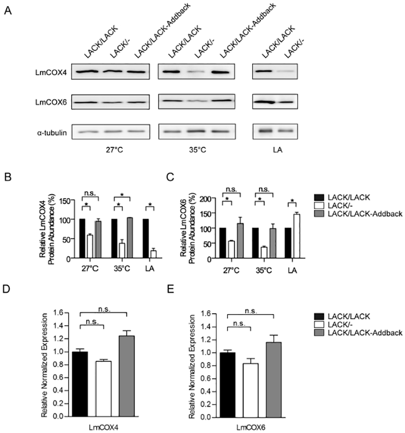
LmCOX4 is decreased in LACK/− L. major incubated at mammalian temperature and in amastigotes.
A. Immunoblot analysis of lysates from L. major lines, as indicated. Left and middle panels: lysates obtained from parasites incubated for 4 days at 27°C or 35°C as previously described (Choudhury et al., 2011). Right panel: L. major lesion-derived amastigotes (LA), as indicated, were purified from the footpads of infected BALB/c mice. The immunoblots were probed with antisera raised against trypanosomatid COIV (LmCOX4), trypanosomatid COVI (LmCOX6) and α-tubulin as denoted.
B and C. Quantification of LmCOX4 and LmCOX6 band intensities, respectively, from LACK/LACK, LACK/− and LACK/LACK-Addback lines at different conditions, as denoted in the figure. Averaged band intensities from three to four immunoblots were analyzed using ImageJ, normalized to α-tubulin and displayed as a percentage of the intensity of its corresponding band in LACK/LACK L. major. Data are displayed as a mean; error bars represent standard error of the mean (SEM) [n = 3 for LmCOX4 (Fig. 2B) and n = 4 for LmCOX6 (Fig. 2C)].
D and E. Reverse transcriptase quantitative PCR analysis of LmCOX4 and LmCOX6 expression at 35°C respectively. Relative expression was normalized to α-tubulin. Data are displayed as a mean; error bars represent SEM (n = 3). *P< 0.05. Two independently derived clones of each parasite line were used in these experiments.
