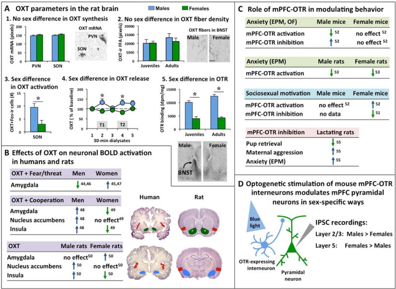
Figure 2. Sex differences in the oxytocin (OXT) system in the rat brain and sex-specific effects on brain activation and behavior by the OXT system.

(A) Analysis of the rat OXT system reveals sex differences in some, but not all OXT parameters: 1. No sex differences are found in OXT mRNA expression in the paraventricular nucleus (PVN) and supraoptic nucleus (SON) of the hypothalamus in adult rats [adapted from 41]. The autoradiogram depicts OXT mRNA expression (black) in the left PVN and left SON in a 16-μm coronal brain section of an adult male rat [adapted from 41]. 2. No sex differences are found in OXT-immunoreactive (OXT-ir) fiber density (expressed as fiber fractional area or FFA) in the bed nucleus of the stria terminalis (BNST) of juvenile and adult rats [adapted from 16]. Photomicrographs depict OXT-ir fibers in the BNST of a male and female adult rat [adapted from 16]; Scale bar indicates 100 μm. 3. Juvenile male rats have more Fos-positive OXT-ir neurons in the SON than females [38]; Fos is an immediate early gene used as marker for neuronal activation. 4. Adult male rats show higher extracellular OXT release (calculated as percentage of baseline OXT release) in the posterior BNST compared to females during both trials of the social discrimination tests in which the rats were exposed to an unfamiliar sex-matched juvenile rat during trial 1 (T1) and the same previously encountered unfamiliar juvenile rat (now familiar) along with a second unfamiliar sex-matched juvenile rat during trial 2 (T2) [adapted from 39]. 5. Juvenile and adult male rats show denser OXT receptor binding in the posterior BNST compared to females [adapted from 18]. Autoradiograms show representative OXT receptor binding in the posterior BNST of an adult male and adult female rat [adapted from 18]. (B) Intranasal application of OXT in humans and intracerebroventricular administration of OXT in rats induce sex-specific blood-oxygen-level dependent (BOLD) activation of the amygdala, nucleus accumbens, and insula [44–50]. In the human studies, men and women were exposed to fearful and/or threatening images or scenes (Fear/threat) or were exposed to an interactive social game (the Prisoner’s Dilemma Game) to examine cooperative interactions (Cooperation). Images depict coronal sections of the human brain (source: https://msu.edu/user/brains/brains/human/) and rat brain [source: 64]. Colors in the coronal brain section depict the amygdala (blue), nucleus accumbens (green), and insula (red). (C) Studies in mice show that the OXT receptor (OTR) in the medial prefrontal cortex (mPFC) regulates anxiety-related behavior and sociosexual motivation in sex-specific ways [51, 52]. In contrast, the mPFC-OTR in rats regulates anxiety-related behavior similiary in males and females [53]. Moreover, the mPFC-OTR is involved in maternal care (pup retrieval), maternal aggression, and anxiety in lactating rats [55]. (D) Studies in mice have shown that in vitro optogenetic stimulation of OTR-expressing interneurons induces a stronger inhibitory postsynaptic current (IPSC) in layer 2/3 pyramidal neurons (important for intra-mPFC connectivity) in males compared to females and a stronger IPSC in layer 5 pyramidal neurons (important for output to subcortical regions) in females compared to males [52]. * p<0.05 versus females.
