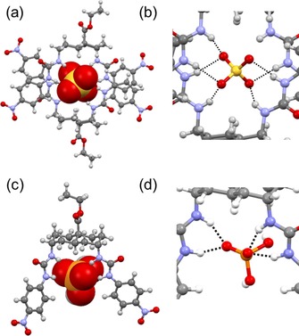
Figure 4.

a) X‐ray crystal structure of (1)2⋅SO4 2−. The transporter is shown in ball‐and‐stick mode, and the anion in CPK mode. b) A closer view of the H‐bonds around the sulfate anion in the dimer. The Me4N+ cation and solvent molecule are omitted for clarity. c) DFT model of 1⋅H2PO4 −. d) A closer view of the H‐bonds around the dihydrogenphosphate anion. H‐bonds are shown as black dashed lines.
