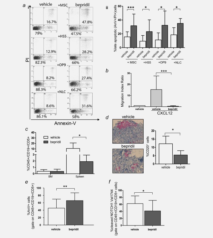
Figure 6.

Bepridil overcomes CLL microenvironment protection, reduces cell migration and exerts antitumor activity in vivo. (a) Representative dot plots (i) of Annexin‐V/PI analysis performed on CLL cells treated with bepridil in the presence of primary mesenchymal cells (MSCs), HS‐5, OP9 cell lines and autologous nurse like cells (NLC). Statistical graphs of late apoptosis (ii) calculated relative to untreated controls (MSCs N = 34; HS‐5 N = 8; OP9 N = 14; NLC N = 6). Resulted are the mean ± SD of all the samples analyzed, *p < 0.05, ***p < 0.001 according to Student's test. (b) Statistical graphs of CLL cells chemotaxis toward CXCL12 (200 ng/mL) in presence or absence of bepridil. Data represent the mean ± SD of three‐independent experiments, each performed in triplicate. ***p < 0.001 according to Student's test. (c) Quantitative recovery of human CD45+/CD19+/CD5+ cells in the bone marrow and spleen of NSG mice evaluated by flow cytometry analysis. (d) Immunohistochemistry analysis and quantification of CD20+ human CLL cells engrafted in the spleen of vehicle or bepridil‐treated mice (original magnification 100×). (e) Percentage of apoptosis estimated by Annexin V staining in CD45+/CD19+/CD5+ cells recovered from the spleen of treated (N = 21) vs. untreated mice (N = 22). Data are mean ± SD. **p < 0.01. (f) Bar graphs show the means ± SD of cleaved‐NOTCH1‐Val1744 fluorescence on splenic CD45+/CD19+/CD5+ CLL cells of 16 bepridil‐treated vs. 17 untreated mice. *p < 0.05.
