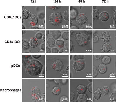
Figure 4.

Localization of antigen storage in APC subsets in vivo. BL/6 mice were injected with Ab i.v. followed by OVA (Alexa Fluor 647 labeled) injection after 30 min. Four APC subsets were sorted at different time points according to the markers described in Fig. 3A and antigen presence in live cells was visualized by confocal microscopy. Data are from a single experiment representative of three experiments with two mice per experiment.
