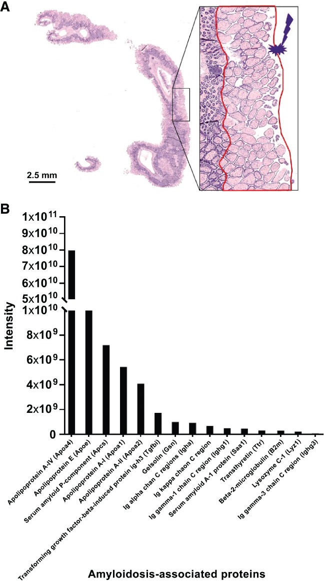
Figure 3.

Characterization of the type of amyloidosis using laser microdissection‐based microproteomics. (A) Congo red‐stained section of the intestine of a PACAP KO mouse. The regions surrounded in red in the inset correspond to the laser‐microdissected one from a serial section. (B) Intensities of amyloidosis‐related proteins identified in the sample. The high intensity of Apoa4, Apoe, Apcs, and Apoa1 suggests that the amyloidosis type is AApoA4.
