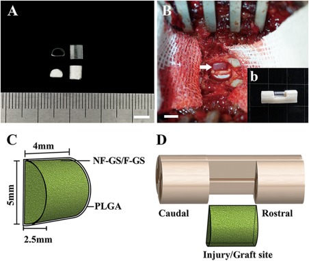
Figure 2.

Scaffold preparation and transplantation. A: Showing two PLGA tubes (top) with a semi‐diameter of 2.5 mm and two gelatin sponge D‐shaped scaffolds (bottom). B: Showing a scaffold transplanted into the hemisection site (arrow, b) of spinal cord. C: Schematic diagram of a scaffold. D: grafted a scaffold in the hemisection site of spinal cord. Scale bars = 5 mm in (A) and (B).
