Figure 7.

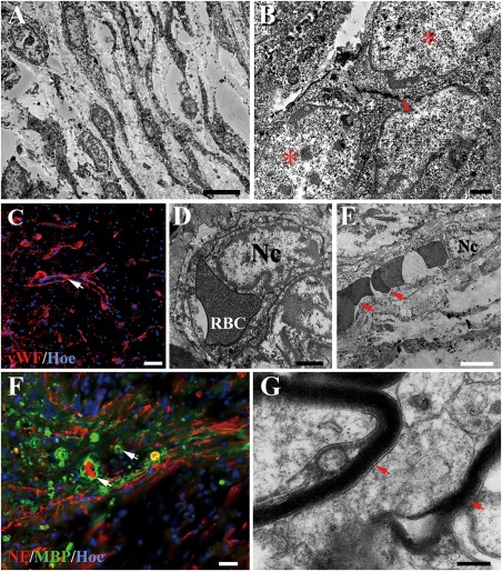
Cell migration and revascularization in the injury/graft site of spinal cord in the NF‐GS group. A: TEM shows many cells appear to migrate into NF‐GS implant. B: A neuron‐like cell (asterisk) containing an aggregation of flattened “synaptic” vesicles (arrow) appears to make a synapse‐like contact associated with another neuron‐like cell (asterisk) under the electron microscopy. C: Note the presence of vascular profiles in the injury/graft site in confocal microscopy, the white arrow indicates the vascular walls. Blood capillaries in transverse (D) or longitudinal (E) section are identified by the lining endothelial cells showing an irregular nucleus (Nc). Note the endothelial cell is invested by a layer of basal lamina (D). Erythrocytes (RBC, arrows) are present in the vascular lumen (E). F, G: Some unmyelinated nerve fibers (NF positive) and a few profiles of myelinated nerve fibers (arrows) were found in the injury/graft site. Scale bars = 5 μm in (A); 2 μm in (B); 50 μm in (C) and (D); 1 μm in (E); 25 μm in (F); 1 μm in (G).
