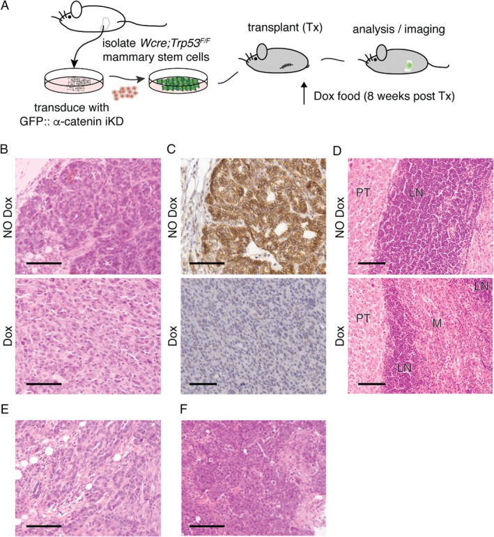
Figure 5.

Inducible loss of α‐catenin leads to invasive mammary carcinoma in a conditional breast cancer progression mouse model. (A) Transplantation‐based models to study breast cancer progression. Schematic overview of the mammary stem cell (MSC) transplantation (Tx) system. Unswitched MSCs were harvested from conditional prepuberal female WAPcre;Trp53 F/F donor mice. After transduction with GFP‐tagged inducible knockdown (iKD) lentiviruses, WAPcre;Trp53 F/F::α‐catenin iKD MSCs were orthotopically transplanted into the cleared fat‐pad of recipient nude 8‐week‐old female mice. Induction of the second tumor suppressor ‘hit’ (loss of α‐catenin) was induced by feeding the mice doxycyclin (Dox)‐containing chow, and mice were monitored for tumor formation and progression. (B, C) Loss of α‐catenin leads to a switch from non‐invasive to invasive carcinoma. Experimental set‐up was as in A, and α‐catenin was inactivated in WAPcre;Trp53 F/F::α‐catenin iKD with doxycyclin 8 weeks post‐transplantation. Tumors were harvested, subtyped using hematoxylin/eosin (HE) (B), and stained for α‐catenin using immunohistochemistry (brown). Upper panels represent control mice (NO Dox); lower panels depict tumors from Dox‐fed mice (Dox). Note the marked acquisition of invasive growth (B and C top versus bottom panels) and the loss of α‐catenin in the Dox‐treated animals (C, bottom panel). (D) Loss of α‐catenin leads to invasion of blood vessels and regional lymph nodes. Tumors from a control (NO Dox, upper panel) and Dox‐fed mouse (Dox, bottom panel) that depict strong differences in local invasion of the inguinal lymph node (LN) are shown. PT = primary tumor; M = metastasis. (E, F) Loss of α‐catenin induces occasional lobular‐like phenotypical features. A representative example of trabecular ducto‐lobular growth patterns (E) and a lesion resembling solid‐type mouse ILC (F) are shown. Scale bars = 50 μm.
