
Figure 2.
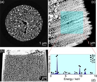
2D perspective of the internal catalyst structure acquired by: (a) PXCT—orthographic slice through the phase contrast tomogram following reconstruction; (b) FIB‐SEM‐CT—secondary electron image of a typical surface exposed during cutting; (c) STEM image of CeOx/np‐Au and (d) corresponding EDX spectrum acquired from the shaded area.