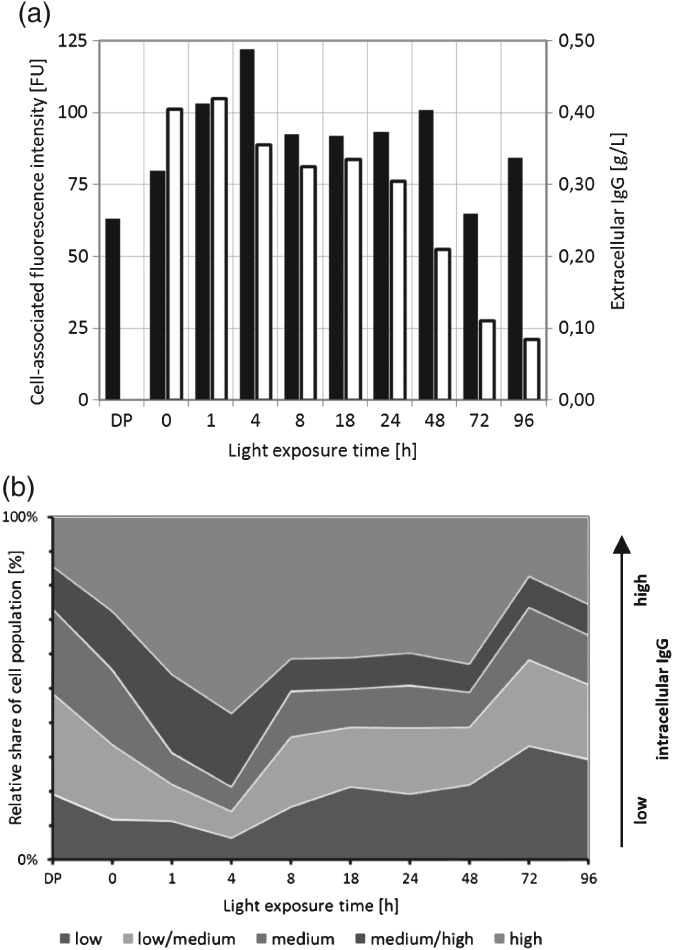
Figure 7.

Analysis of IgG formation in CHO shake flask cultures in light‐irradiated CDPM. (a) Intracellular IgG was detected via fluorescence‐immunostaining in fixed and permeized cells with subsequent flow cytometric analysis (mean cell‐associated fluorescence intensity; closed bars), extracellular IgG was quantified by HPLC (open bars). (b) Proportional composition of the CHO cell population at different light exposure times. For each time point, cells were stratified according to the intracellular IgG level (high/medium‐high/medium/medium‐low/low intracellular IgG staining).
