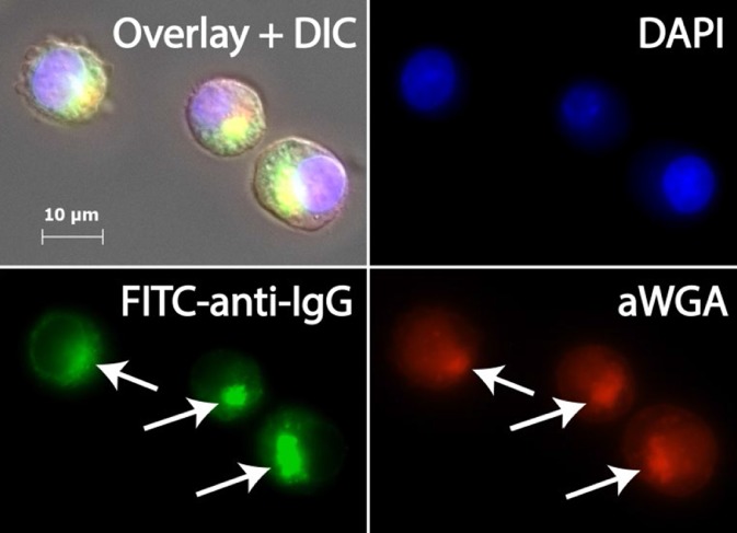
Figure 8.

Fluorescence micrograph of intracellular IgG localization in CHO cells. Cells from shake flask cultivation (0 h LET, end of exponential phase) were fixed, permeized, and stained with the FITC‐labeled anti‐IgG antibody (FITC‐anti‐IgG). Alexa594‐conjugated wheat germ agglutinin (aWGA) was used as a Golgi marker, and nuclei were labeled with a DNA‐specific stain (DAPI). Note the high degree of co‐localization between IgG and Golgi compartments (arrows), which is clearly visible in the morphological overlay with differential interference contrast (Overlay + DIC).
