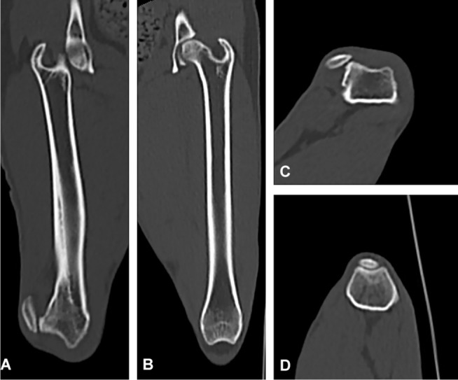
Figure 4.

CT scan of a dog with unilateral lateral patellar luxation.
Notes: (A) Frontal view of the femur showing dislocation of the patella laterally and distal femoral bowing. (B) Normal. (C) Transverse view showing abnormal shape of the trochlear groove and lateral dislocation of the patella. (D) Normal.
