
Fig. 1.
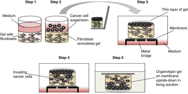
Schematic representation of the workflow of an organotypic Invasion assay. Step 1: Embed fibroblasts in gel and seed in 24-well dish. Step 2: Cancer cells are seeded in a single-cell suspension on top of the gel. Step 3: Once the cells have adhered, remove the medium and lift the remodeled gel onto gel-coated Nylon filter on a metal bridge. Coat the cancer cells with a thin layer of gel. Step 4: Feed with complete medium up to the Nylon filter. Incubate at 37 °C, 5% CO2 for 5 days to allow for cancer cell Invasion. Step 5: Terminate assay by fixing organotypic gels. Process gels for H&E staining