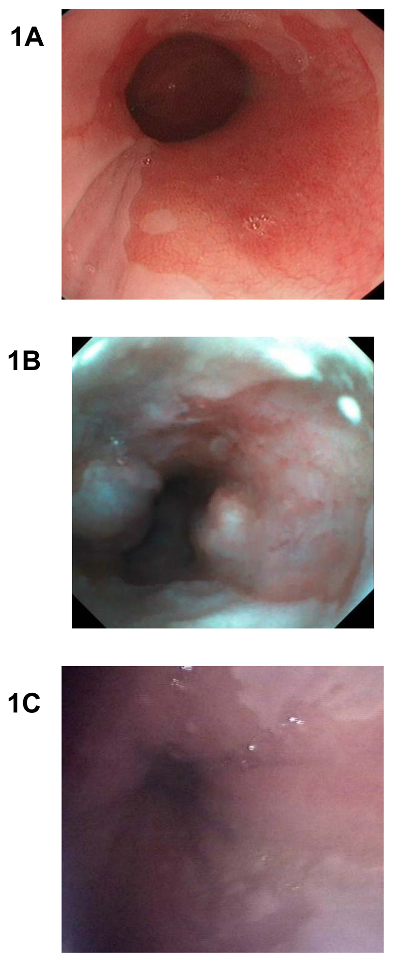
Fig. 1. Endoscopic diagnosis of Barrett’s esophagus with conventional per-oral and office-based transnasal endoscopy.
(A) High resolution white light endoscopy. Barrett’s esophagus appears as salmon red coloured mucosa and normal oesophagus in pale pink. (B) Transnasal EG scan endoscopic view of a short segment of Barrett’s esophagus. (C) Transnasal endosheath endoscopic diagnosis of Barrett’s. This technology also allows biopsies for histological confirmation.

