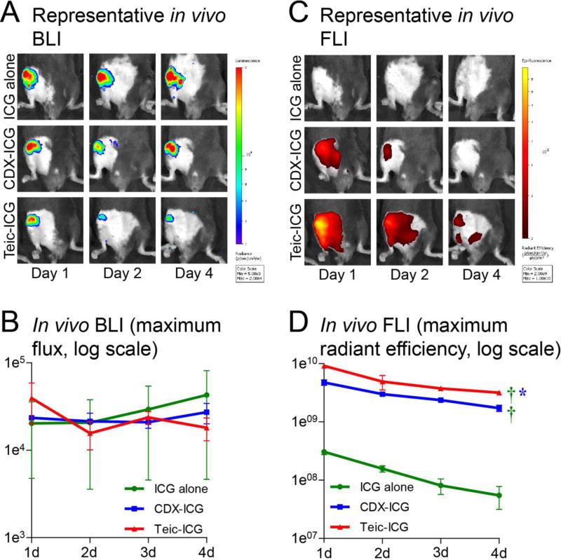
Figure 1. In vivo bioluminescence (BLI) and fluorescence imaging (FLI).
Mice underwent implantation of a Kirschner-wire with direct inoculation of bioluminescent S. aureus into the right knee. Seven days after surgery, three hybrid fluorescent / photoacoustic tracers were injected intravenously: indocyanine green (ICG) alone, or tagged to β-cyclodextrin (CDX-ICG) or teicoplanin (Teic-ICG). (A) Representative in vivo bioluminescence images, with signal indicative of infection localized to the surgical site. (B) Overall bioluminescence of the three tracers. No significant difference in signal was noted. (C) Representative in vivo fluorescence images, with localization to the surgical site in Teic-ICG and somewhat in CDX-ICG but not in ICG alone. (D) Overall fluorescence of the three tracers. Teic-ICG (P=0.0087) and CDX-ICG (P=0.0046) were both significantly higher than ICG alone. Teic-ICG was also significantly higher than CDX-ICG (P=0.047).

