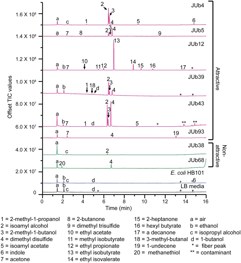
Fig 2. Gas chromatography-mass spectrometry of headspace of natural bacterial isolates, E. coli HB101 and LB media control.
Overnight liquid cultures of bacteria were spotted on NGM agar plates (OD600 = 10) and incubated for 1 hour, then NGM agar squares with bacterial suspension were placed inside a GC-MS glass vial for five hours. A SPME fiber was inserted into the vial containing the bacteria to sample the volatile compounds in the headspace. Top, representative total ion chromatograms (TIC), from top to bottom: JUb4, JUb5, JUb12, JUb39, JUb43, JUb93, JUb38, JUb68, E. coli HB101and LB media control. Peaks present in bacterial samples, but not LB media, are numbered. Peaks present in LB media are labeled with letters. One asterisk indicates “fiber peaks,” volatile siloxanes released by the SPME fiber; two asterisks indicate contamination because also present in fiber blank control run prior to JUb43 sample analysis. Bottom, volatile organic compounds (VOCs) corresponding to labelled peaks. Peaks were identified tentatively with NIST 11 (National Institute of Standards and Technology) mass spectral library and confirmed with known standards. Retention times of VOCs in samples JUb43, JUb38, and JUb68 are shorter by approximately 0.1 minute compared to other samples analyzed because a small, contaminated segment of the column was removed from the inlet. See sample retention times in S1 Table. All bacterial samples were prepared and analyzed two or more times on different days.

