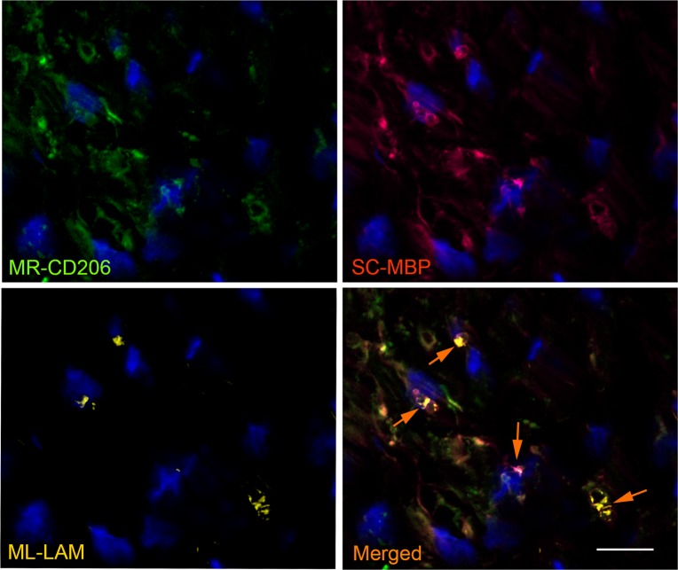
Fig 10. M. leprae infected Schwann cells express CD206 in leprosy nerve lesions.
Nerves biopsies were labeled with antibodies for mannose receptor CD206 (green image), for the SC-specific marker MBP (red image) and for M.leprae (anti-LAM; yellow image). Nuclei were labeled with DAPI (blue). The serial section of a leprosy patient nerve biopsy was analyzed by fluorescence microscopy. The images (representative of two patients) show the expression profile of the CD206 mannose receptor, the MBP SC marker and the location of M. leprae. The merged image shows CD206/MBP/LAM co-staining in SCs (white arrows). Scale bar, 20μm.

