
Figure 3.
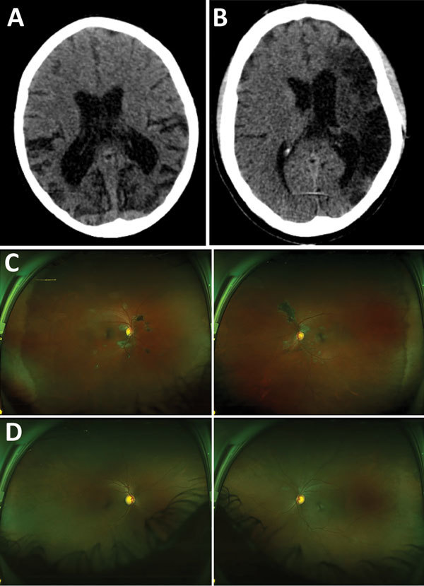
Representative nonenhanced computed tomography (CT) brain scans and composite scanning laser ophthalmoscope fundus images of 2 Ebola virus disease survivors attending a joint neurologic and psychiatric clinic in Sierre Leone. A) Patient no. 37, female, age 12. CT of brain shows disproportionate parietal and temporal lobe atrophy. B) Patient no. 25, male, age 42. CT of brain shows extensive gliosis within the left middle cerebral artery territory reflects an old infarct with ex-vacuo dilatation of left lateral ventricle due to hemispheric volume loss. C) Patient no. 12, age 40. Retinal imaging shows left and right eye, with extensive bilateral peripapillary pale retinal lesions with pigmentation of larger lesions. Lesions appear to spare the fovea. Visual acuity was 20/25 (right) and 20/20 (left) (24). D) Patient no. 25, male, age 42. Retinal imaging shows left and right eye, with peripapillary pale retinal lesions. Visual acuity was 20/25 in both eyes (25).