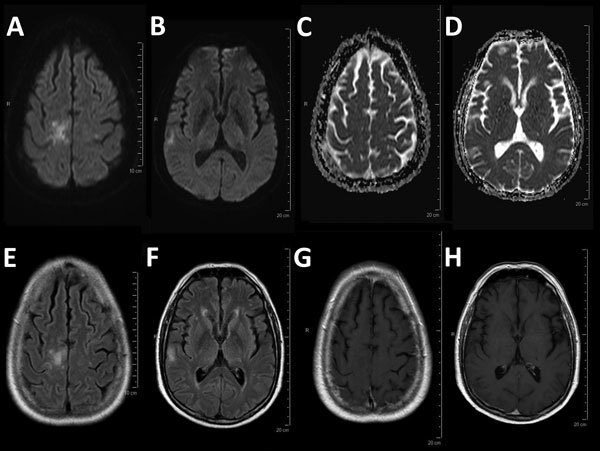
Figure.

Magnetic resonance imaging for a 54-year-old man with progressive multifocal leukoencephalopathy after treatment with nivolumab, showing typical multifocal lesions: diffusion weighted imaging hyperintensity (A, B) with no decrease in the apparent diffusion coefficient (C, D), corresponding patchy corticosubcortical hyperintensities on fluid-attenuated inversion recovery image (E, F) without enhancement on T1-weighted imaging after administration of gadolinium (G, H).
