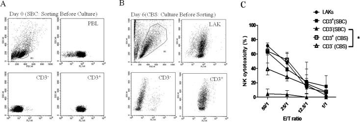
Fig. 1.
A: CD3+ (SBC) (■) and CD3– (SBC) (▲) cells sorted from PBLs and cultured separately with IL-2 for 6 days before perfoming the NK killing assay. B: PBLs stimulated with IL-2 (2500 U/ml) for 6 days, and CD3+ (CBS) (□) and CD3− (CBS) (△) cells sorted for a NK killing assay. C: Killing activities are shown in Fig. 1C

