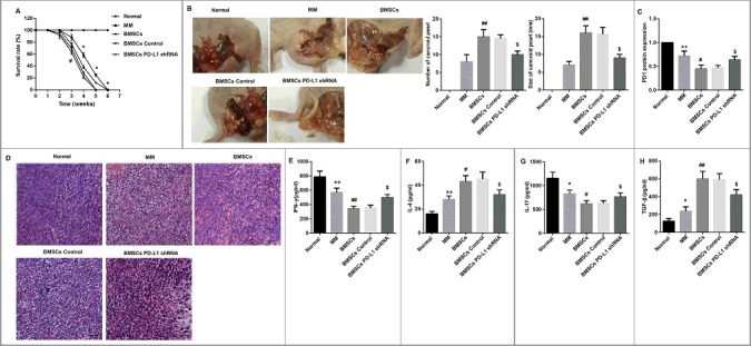
Figure 6.

Effects of BMSCs and PD-L1 shRNA in MM model mice. (A) 6-week survival rates of mice in each group was drawn (for each group, n = 12). (B) After 6 weeks of feeding, the cancroid pearls in the abdominal cavity of mice were separated. Representative photographs of typical cancroid pearls in the abdominal cavity of mice were shown. The number and size of cancroid pearls in the abdominal cavity were shown as column graphs. (C) The tumor tissues from abdominal cavity of mice in each group were isolated, partly for protein extraction for PD-1 evaluation by Western blot (for each group, n = 3), (D) partly for HE staining for observation of pathological changes and inflammation infiltration (for each group, n = 4; Scale bar: 20 μm), and partly for tissue homogenate to detect (E) IFN-γ, (F) IL-4, (G) IL-17, and (H) TGF-β by ELISA (for each group, n = 5). *P<0.05, **P<0.01 vs. Normal. #P<0.05, ##P<0.01 vs. MM. &P<0.05 vs. BMSCs Control.
