
Figure 1.
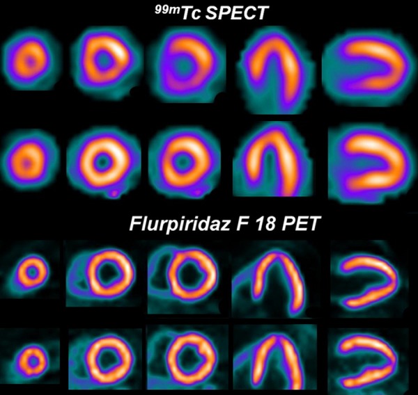
99mTc SPECT images (upper rows) and 18F-flurpiridaz PET images (lower rows) from a patient with normal coronary arteries. A false positive reversible inferior defect is present on the 99mTc SPECT images due to shifting soft-tissue attenuation. The 18F-flurpiridaz PET study, however, provided superior image quality and was normal.