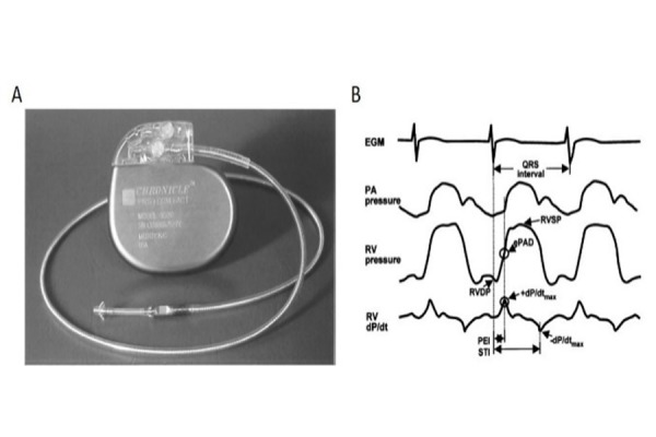
Figure 1. Right ventricular device (Chronicle).

A: Right ventricular implantable device (Chronicle) with the pressure sensor at the distal tip of the single right ventricular lead; B:
Representative waveforms obtained from the device: electrocardiogram, right ventricular (RV) pressure, pulmonary artery (PA) pressure and right ventricular dP/dt, ePAD: estimated pulmonary artery diastolic pressure; +dP/dtmax: maximum positive dP/dt; -P/dtmax: maximum negative dP/dt; PEI:pre-ejection interval; STI: systolic time interval. Reproduced with permission of [22]
