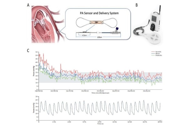
Figure 2. Pulmonary pressure sensor device (CardioMEMS).

A: CardioMEMS sensor and delivery catheter and schematic representation of the implanted sensor; B: external electronic monitoring unit connected with antenna used to calibrate the device and to take readings of hemodynamic data; C: representative haemodynamic data provided by the device, including pulmonary rtery pressure waveforms (systolic, mean and diastolic) and a pressure trend graph. Reproduced with permission of [24].
