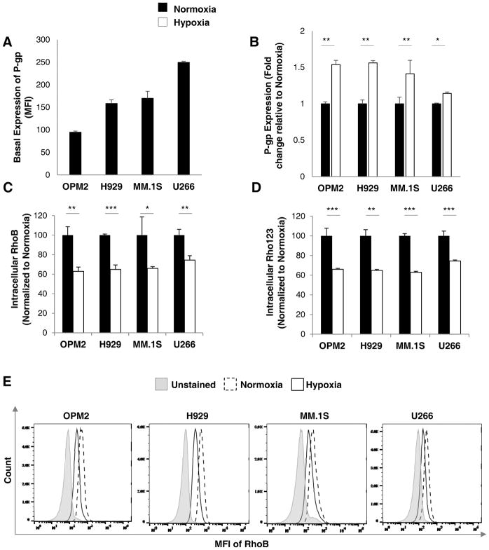
Figure 2. Hypoxia increases P-gp protein expression and activity in MM cells.
(A) Basal expression of P-gp protein across MM cell lines (OPM2, H929, MM.1S and U266) cultured in normoxia (21% O2; 24 hours) analyzed by flow cytometry and demonstrated as MFI. (B) The expression of P-gp protein in normoxia (21% O2; black bars) and hypoxia (1% O2; white bars) for 24 hours analyzed by flow cytometry demonstrated as fold change and normalized to normoxia. P-gp activity shown as intracellular RhoB (C) and Rho123 (D) content in MM cell lines (OPM2, H929, MM.1S and U266) cultured in normoxia and hypoxia for 24 hours measured by flow cytometry and depicted as MFI normalized to unstained cells, relative to normoxic cells. (E) Histograms demonstrating RhoB in normoxia and hypoxia in MM cell lines. Results are shown as mean ± standard deviation (s.d.); the statistical significance was assessed by student t-test and considered significant for values * p<0.05, ** p<0.01 and *** p<0.001.

