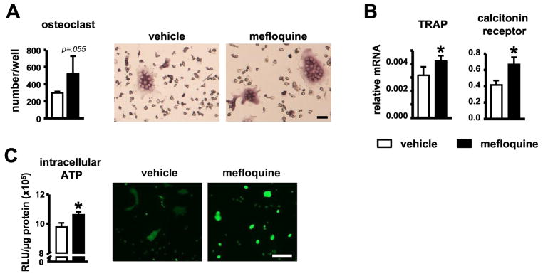
Fig. 5. Increased serum bone resorption markers in young and old mice, and osteoclast differentiation in vitro induced by mefloquine.
(A) Osteoclast number and (B) genes associated with osteoclast differentiation in cultures of non-adherent bone marrow cells treated with M-CSF and sRANKL, measured by qPCR and corrected by GAPDH (n=4). Representative images of in vitro generated osteoclasts are shown. Scale bars indicate 10μm. (C) ATP levels were measured in osteoclast lysates (intracellular ATP, n=3) after 7 days of differentiation using a luciferin-luciferase kit. Mature osteoclasts treated with vehicle or 1mM mefloquine for 24h were stained using 100μM quinacrine and visualized under fluorescence microscope (n=3). Scale bars indicate 100μm. Bars represent mean ± s.d. *p<0.05 versus vehicle treated group, by t-test.

