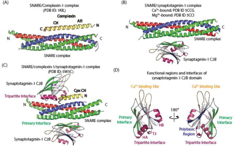
Figure 2. Atomic-resolution structures of SNARE/complexin-1, SNARE/synaptotagmin-1, and SNARE/complexin-1/synaptotagmin-1 complexes.
(A) Crystal structure of the pairwise complex between complexin-1 (yellow) and the SNARE complex (synaptobrevin-2, blue; SNAP-25, green, syntaxin-1A, red) [63] (PBD ID 1KIL). (B) Superposition of the Ca2+ and Mg2+-bound crystal structures of the pairwise complex between the SNARE complex (synaptobrevin-2, blue; SNAP-25, green, syntaxin-1A, red), and synaptotagmin-1 C2B (gray, green, purple, blue, and gold) [3] (PDB IDs 5CCG and 5CCI). For clarity, only the primary C2B-SNARE interface is shown. (C) Crystal structure of the Ca2+-free tripartite complex between the half-zippered SNARE complex (synaptobrevin-2, blue; SNAP-25, green, syntaxin-1A, red), complexin-1 (yellow), and synaptotagmin-1 C2B (gray, green, purple, blue, and gold) [16] (PDB ID 5W5C). (D) Functional regions and interfaces of the synaptotagmin-1 C2B domain. The colors indicate the loops involved in Ca2+-binding (gold), the primary SNARE-synaptotagmin-1 interface (green), the tripartite SNARE/complexin-1/synaptotagmin-1 interface (purple), and the polybasic region (blue).

