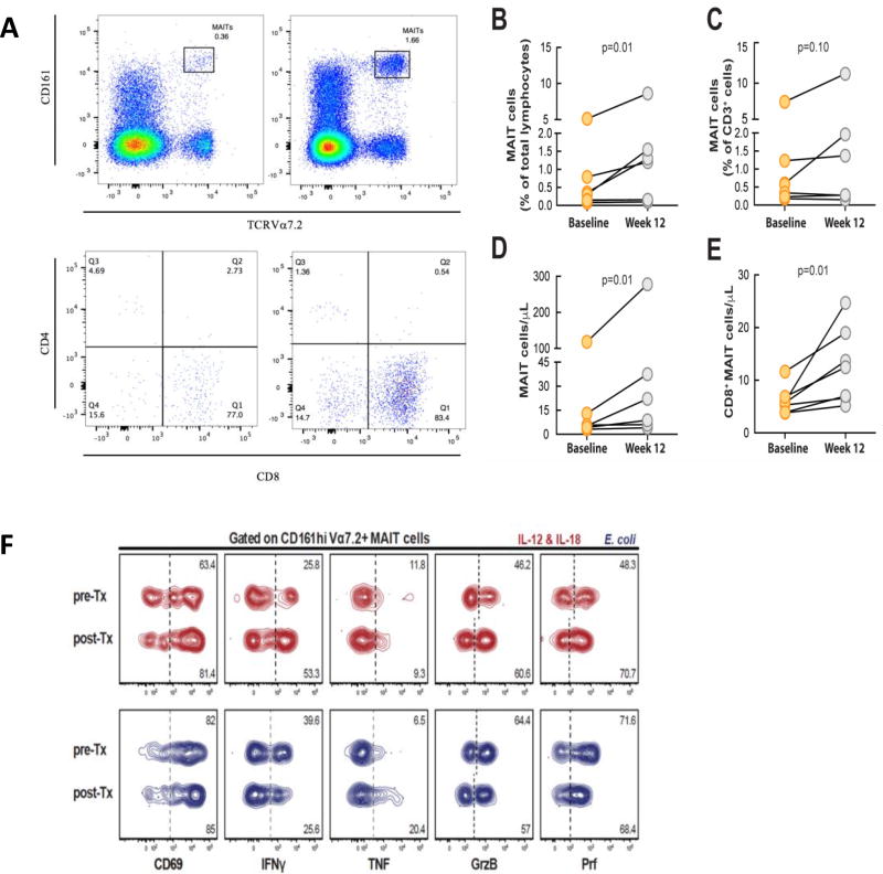
Figure 1. MAIT cell characterization and changes in the MAIT cell population after treatment with IL-7.
(A) MAIT cells identified by Vα7.2 and CD161 staining and flow cytometry. Expression of CD8 and CD4 in MAIT cells. (B) Percentage of MAIT cells among total lymphocytes increased (p = 0.01) after treatment with IL-7. (C) There was no significant increase in the percentage of MAIT cells within the total CD3+ cells. (D) The absolute number of MAIT cells increased significantly after IL-7 treatment (p = 0.01). (E) There was an increase in the absolute number of CD8+ MAIT cells after IL-7 treatment. (F) Function of MAIT cells in a HIV-1 infected patient before and after rhIL-7 treatment after stimulation with E. coli.

