. 2018 Jul 18;38(6):555–562. doi: 10.3343/alm.2018.38.6.555

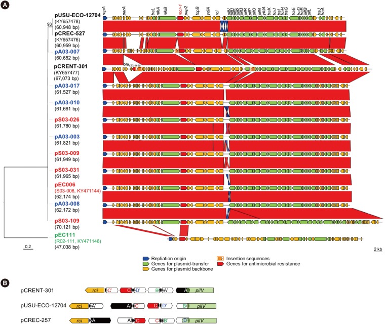
Fig. 1. Comparative analyses of the mcr-1-plasmids from clinical Enterobacteriaceae strains and livestock-origin Escherichia coli strains. (A) Left, molecular phylogeny was conducted by neighbor joining analysis of nucleotide sequences (324 bp) of the replication origin of the mcr-1-plasmids. Multiple sequence alignments were performed with MUSCLE v3.8, and the phylogenetic tree was reconstructed using the distance method implemented in the BioNJ program [34]. Plasmid names are indicated in each taxon along with the size in brackets. Strain name and GenBank accession number are indicated below the plasmid name, if available. Color codes: Black, clinical Enterobacteriaceae strains; blue, E. coli from healthy chickens; red, E. coli from chicken carcasses; and green, E. coli from diseased pig. Right, schematic representation of plasmid structures. The sequence of each plasmid was aligned using BlastN and compared using the Artemis Comparison Tool. Highly-conserved regions (>96% nucleic acid identity) are indicated in red, and moderately conserved regions (>92% nucleic acid identity) are indicated in blue. Open arrows, open reading frames; blue, the replication origin; red, antimicrobial resistance; yellow, plasmid backbone; orange, transposases; and green, plasmid transfer. (B) The yellow arrow indicates the site-specific recombinase rci gene, and the green arrow indicates the pilUs assembly pilV gene. Black arrowheads represent the six 19-bp repeats. Open reading frames in the direction of translation are indicated by arrows.

Fig. 1