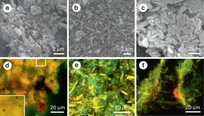
Fig. 2.
Decayed tissues of extant frogs. a–c Scanning electron micrographs of rod-shaped decay bacteria within internal tissues of the torso in Kaloula (a), Xenopus (b) and Osteopilus (c). d–f Fluorescent micrographs of melanosomes (d) and rod-shaped bacteria (e) in the torso, and of rod-shaped bacteria in the thigh (f). Melanosomes appear black in fluorescent images. Bacteria appear green and red. Scale bars, 2 μm (a–c), 20 μm (d–f)

