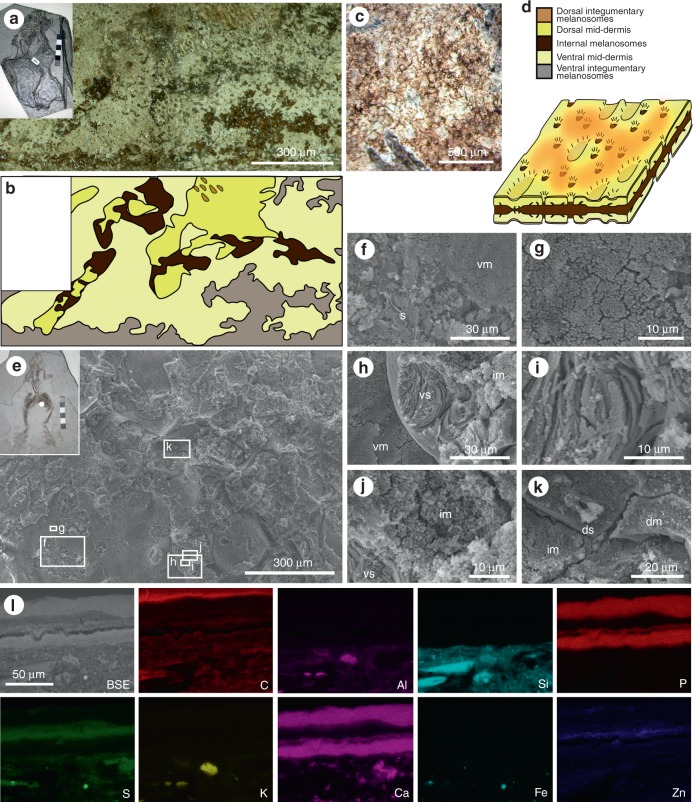
Fig. 4.
Layered soft tissues in the hindlimbs of fossil frogs from Libros. a–b MNCN 63775. Light micrograph of area indicated in inset, showing a plan view of the soft tissues in the thigh. b Interpretative drawing of soft tissue layers present, based on light microscopy. c Patterning in the integumentary melanosome layer. d Three-dimensional schematic illustration of the preserved 3D structure of the layered soft tissues in the Libros frogs, based on data presented in ref.12 and this figure. The terms ‘dorsal’ and ‘ventral’ are for illustrative purposes only. e–l MNCN 63798. e Scanning electron micrograph of soft tissues from the region indicated in the inset. f–k Details of regions indicated in e. f Sediment (s) underlying the ventral melanosome layer (vm). g Detail of ventral melanosomes. h Ventral layer of phosphatized skin (vs) overlain by non-integumentary melanosomes (im) and underlain by ventral melanosomes. i Detail of collagen fibres of phosphatized skin. j Detail of non-integumentary melanosomes. k Dorsal layer of phosphatized skin (ds) overlain by dorsal melanosome layer (dm). l Elemental maps of polished sections through the soft tissues from the thigh. Melanosome layers are defined by C, S and Zn. SE: secondary electron micrograph of region analysed, dm: dorsal melanosome layer, s: sediment, vm: ventral melanosome layer, vs: ventral skin layer. Scale bars, 300 μm (a), and same scale in b, 500 μm (c), 300 µm (e), 30 µm (f, h), 10 µm (g, i, j), 20 µm (k), 50 μm (l)

