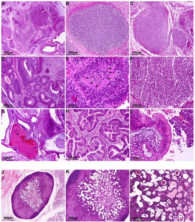
Figure 1.
Histological examination of tumors from hiPSCs and EB cells. Representative microscopic images of Haematoxylin & Eosin-stained sections of (A) hiPSCs derived-teratoma showing the differentiation into all three germ layers; (B,C) mature and immature cartilage (mesoderm); (D,E) primitive neuroectodermal tissue (endoderm) in the form of rosettes with high mitotic activity (arrowheads); (F) cellular primitive stromal tissue; (G) hemorrhage and necrosis (asterisk); (H,I) glandular and columnar epithelial tissue (endoderm); (J,L) a small lymph node associated with fibrosis and edema was found in the site of inoculation of differentiated EBs (20 days).

