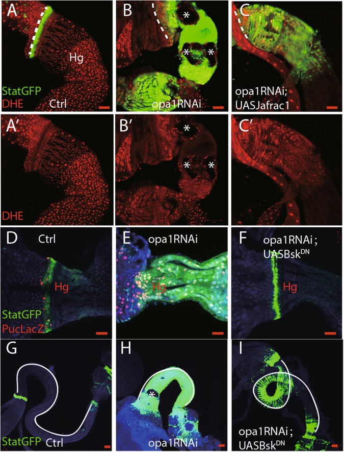
Fig. 5. ROS–JNK pathway contribute to differentiation defects in opa1RNAi hindguts.
a–c DHE staining of freshly dissected hindgut. a′–c′ are DHE channel. Asterisk denotes potentially non-apoptotic cell death in opa1RNAi hindguts. d–f Anti-beta Gal staining against Puc-LacZ in different genotypes. g–i Differentiation index, such as Stat::GFP and gut length, was compared in different conditions. Gut length was delineated out by white lines. Scale bar is 20 µm

