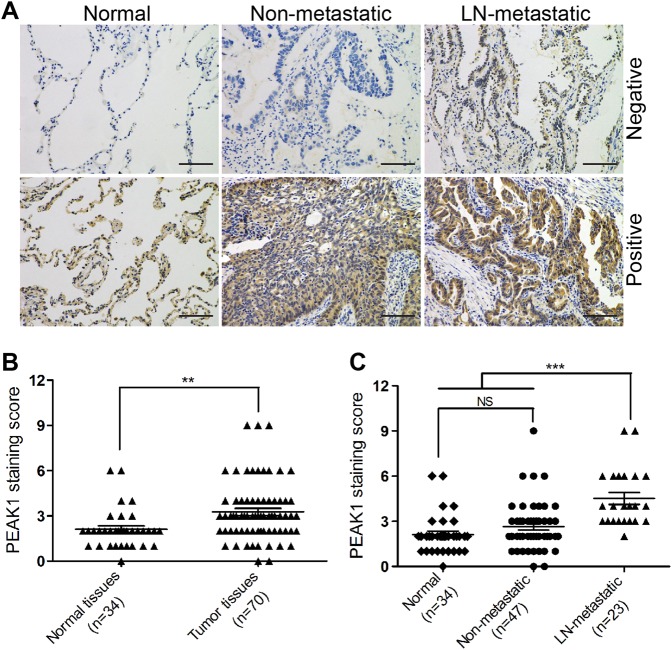
Fig. 4. PEAK1 is markedly upregulated in metastatic lung cancer tissues.
a Immunohistochemistry analysis of PEAK1 expression in lung cancer tissue and adjacent normal tissue. Scale bar, 100 μm. b Immunohistochemical staining of normal and tumor tissues was evaluated by staining scores. c Immunohistochemical staining of normal, non-metastatic tissues and LN-metastatic tissues was evaluated by staining scores. NS no statistical significance; **P < 0.01; ***P < 0.001

