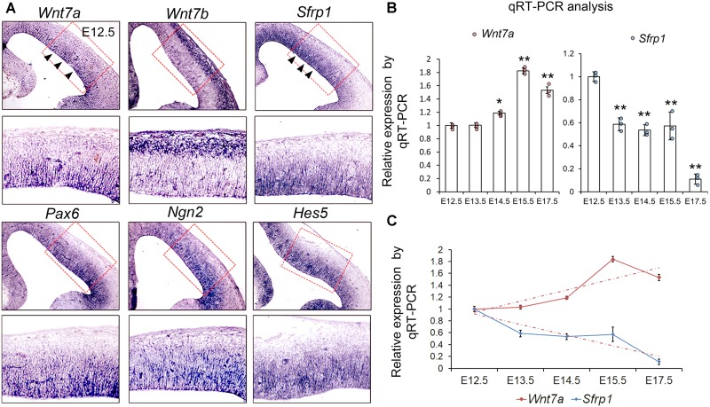
FIGURE 1.
Wnt7a and Sfrp1 are co-expressed in neural progenitors and show opposite expression trends. (A) In coronal sections of mouse E12.5 cerebral cortices, Wnt7a, Sfrp1, Pax6, Ngn2, and Hes5 were expressed in the ventricular zone (arrowheads). Conversely, Wnt7b was expressed in newborn neurons. Red boxes show high power views. (B) qRT-PCR analysis of Wnt7a and Sfrp1 expression levels at different embryonic stages (E12.5, E13.5, E14.5, E15.5, and E17.5). All comparisons were made with that of values at E12.5. Values of histogram represent mean ± SEM, and each dot represents a data point in each biology repeat (n = 3, ∗P < 0.05; ∗∗P < 0.01; unpaired Student’s t-test). (C) Opposite expression trends between Wnt7a and Sfrp1 at different embryonic stages (from E12.5 to E17.5) measured by qRT-PCR.

