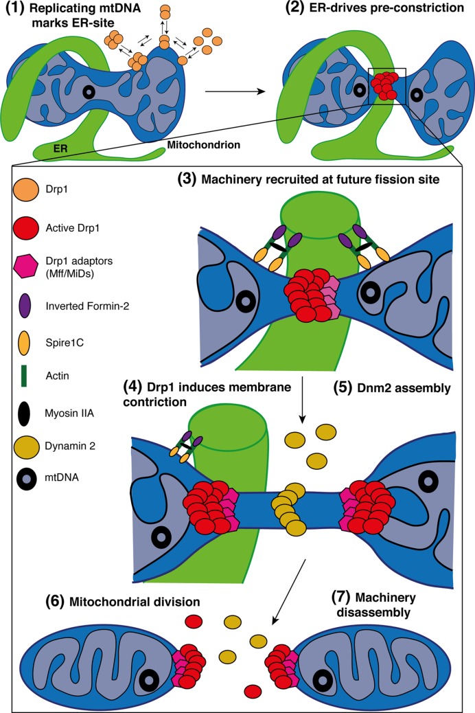
Figure 4. Simplified model for mitochondrial fission in mammals.
Schematic representation of the multi-step processes required for mitochondria division. (1) In the matrix, replication of the mtDNA marks the site for ER-recruitment. In parallel, Drp1 oligomers are in constant balance between the cytosol and mitochondria. In addition, IMM constriction occurs at mitochondria–ER contacts in a Ca2+-dependent process, before Drp1 oligomerization and maturation. (2) Oligomeric forms of Drp1 accumulate at ER-sites where the pre-constriction of the membrane has been initiated. (3) The zoomed area highlights the factors regulating mitochondrial division. The ER-bound INF2 and mitochondrial Spire1C induce actin nucleation and polymerization at mitochondria–ER contact sites. The Myosin IIa may ensure actin cable contraction, providing the mechanical force to drive mitochondria pre-constriction. At these sites, MFF and MiDs recruit Drp1 where it oligomerizes in a ring-like structure and (4) GTP-hydrolysis leads to conformational change, enhancing pre-existing mitochondrial constriction. The composition of the OMM in phospholipids also regulates Drp1 assembly and activity. (5) Then, Dnm2 is recruited to Drp1-mediated mitochondrial constriction neck where it assembles and terminates membrane scission, (6) leading to two daughter mitochondria. (7) The mechanisms of disassembly of the fission machinery following division remain unclear but both adaptors and Drp1 are found at both mitochondrial tips after division.

