
FIGURE 2.
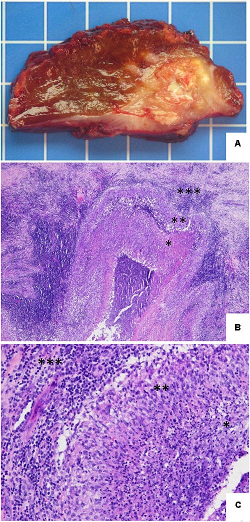
Hepatic histopathology of human brucellosis. (A) Gross section of the hepatic segments showing a large and relatively well-demarcated lesion with some calcifications and a central necrotic and suppurative area. One square = 1 cm2. (B) Hematoxylin and eosin stain. The lesion consists of a large pseudotumoral abscessed lesion with a central necrotic area (∗) surrounded by a serpiginous granulomatous inflammation (∗∗) and associated with abundant peripheral fibro-inflammatory tissue (∗∗∗) (original magnification, ×4). (C) Hematoxylin and eosin stain. Higher power view illustrating the transition between the central suppurative necrosis (∗), delineated by a rim of epithelioid histiocytes arranged in palisade (∗∗), with peripheral fibrous tissue comprising an intense lymphoplasmocytic infiltrate (∗∗∗) (original magnification, ×20).