
Figure 2.
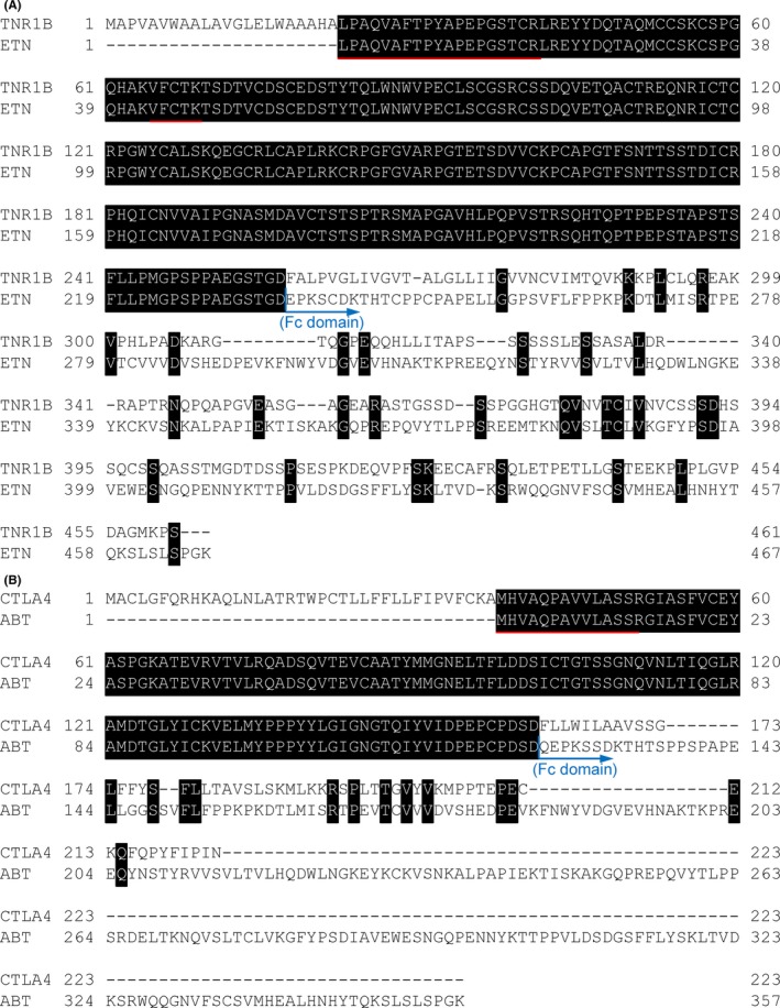
The ClustalW sequence alignment of (A) TNFR (TNR1B) and Etanercept (ETN), and (B) CTLA‐4 (CTLA4) and Abatacept (ABT). The black area shows identical amino acid residues. The red lines show the selected signature peptide of each Fc‐fusion protein. The blue arrow represents the position of the beginning of fused Fc domain