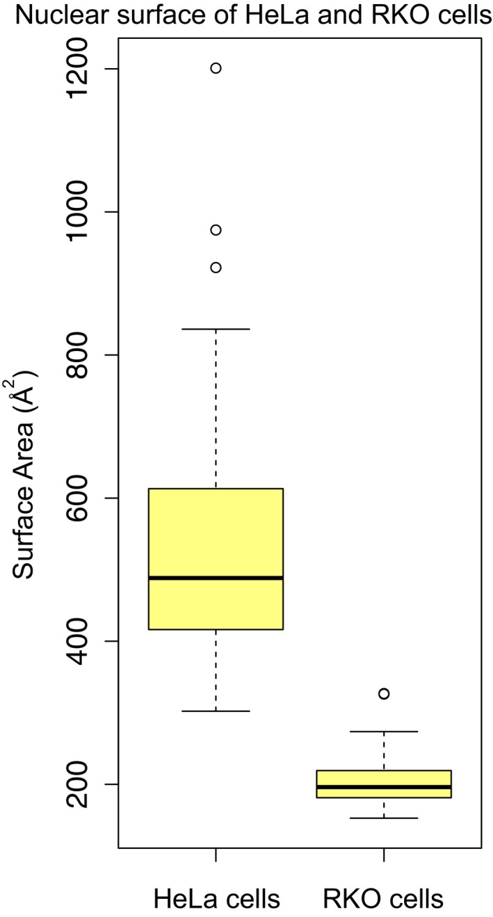
Figure EV3. Comparison of nuclear surface area of HeLa and RKO cells.

Distributions, represented as boxplots, of nuclear surface area (Å2) of 44 HeLa and 41 RKO cells (Ori et al, 2013). HeLa nuclei are significantly larger than RKO nuclei (t‐test P = 2.5 × 10⁻¹⁵). Nuclear surface area was estimated from the radius of isolated nuclei measured in phase contrast images, assuming a spherical shape. Related to Fig 2D and Dataset EV5. Boxplots: the horizontal line represents the median of the distribution, the upper and lower limit of the box indicate the first and third quartile, respectively, and whiskers extend 1.5 times the interquartile range from the limits of the box. Values outside this range are indicated as outliers points.
